Ultrafast OCT optical tomograph for ophthalmic examinations using the technique of tuned lasers (64)

Nicolaus Copernicus University in Toruń

  • biomedical studies
  • OCT angiography
  • ophthalmological studies
  • optical studies

Contact person :

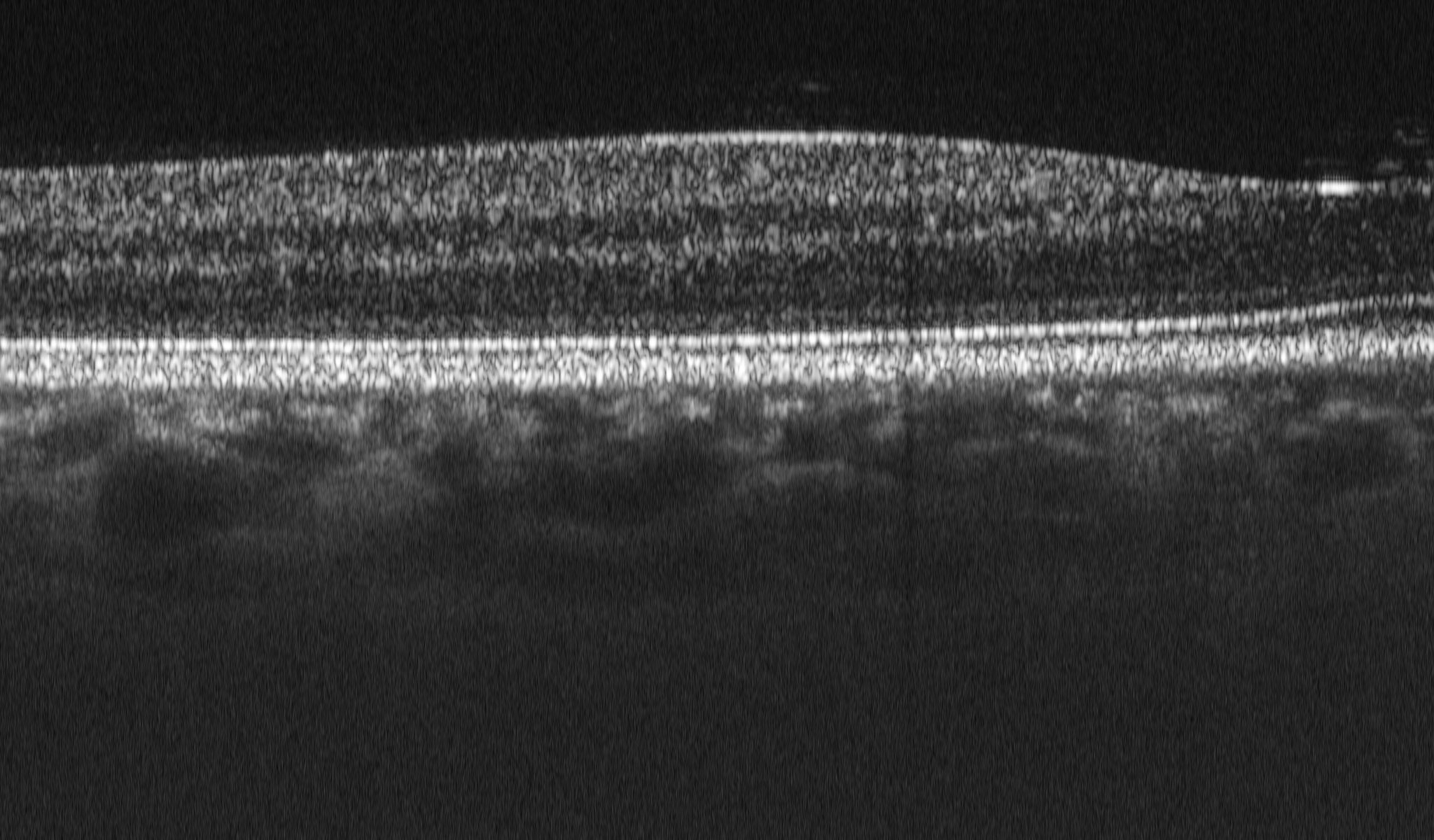
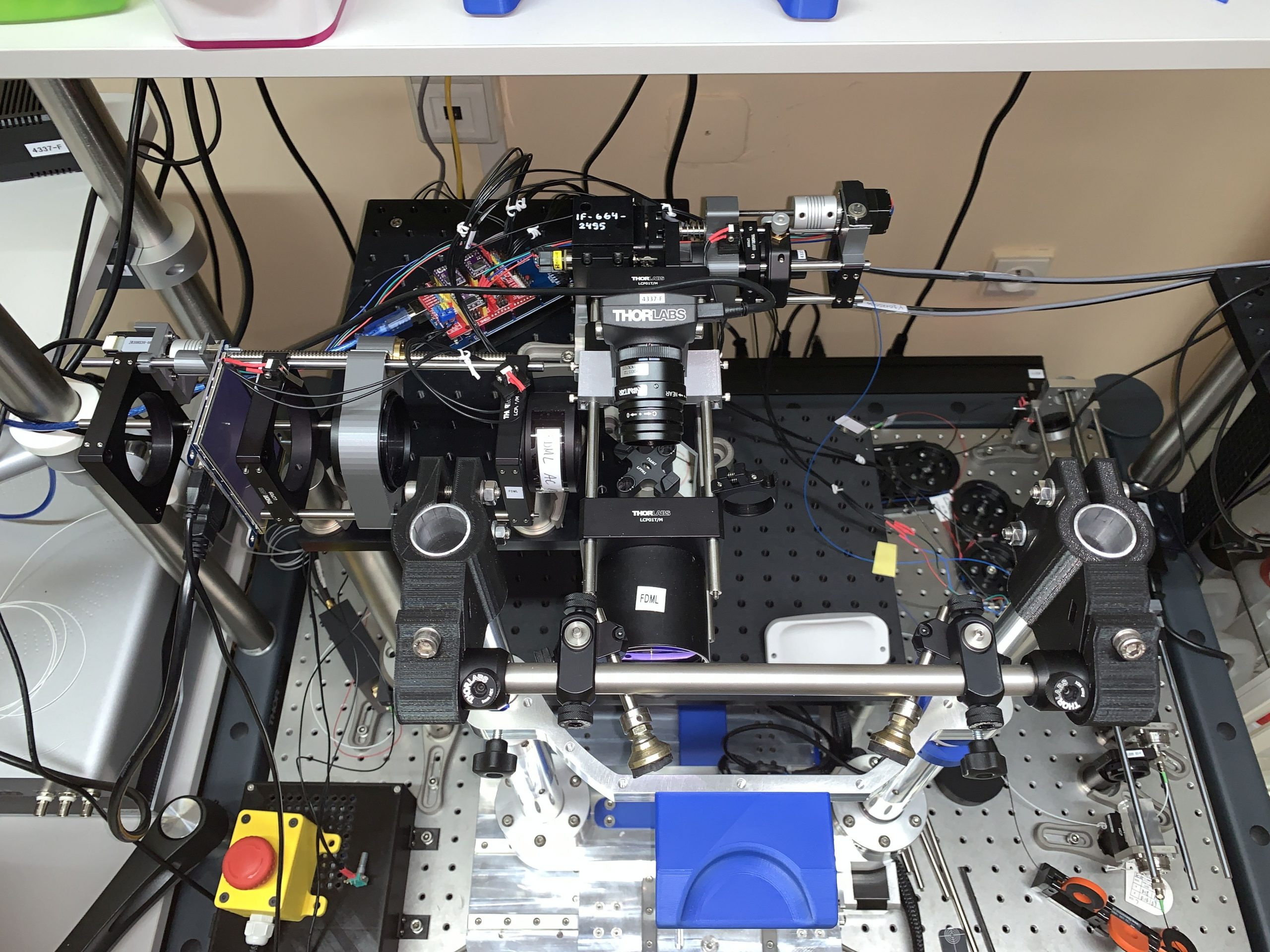
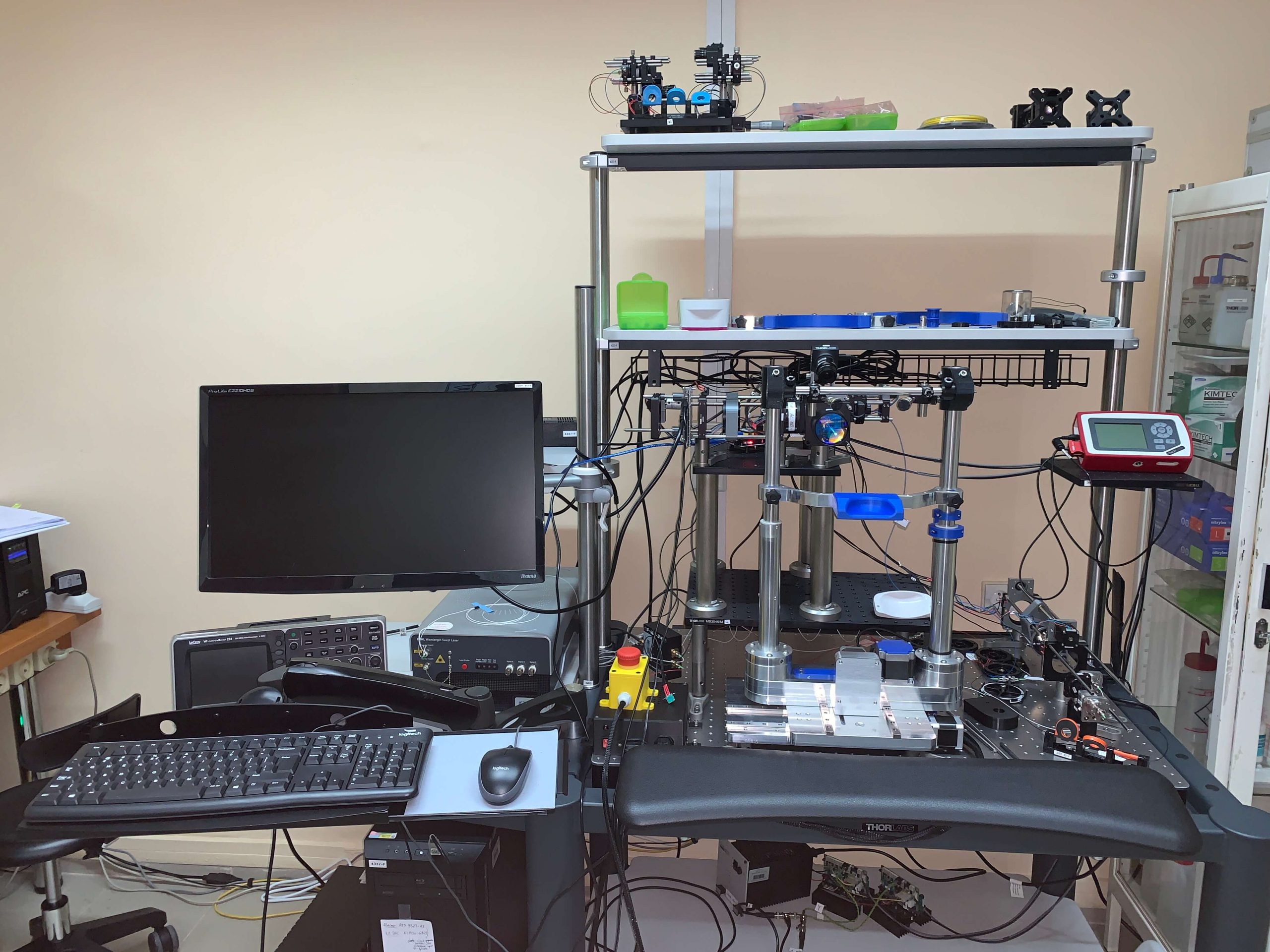
The OCT optical tomograph is designed for examining the fundus of the human eye, i.e. the retina, the optic disc and the uveal membrane. The device operates in the OCT technique in the Fourier field with the use of tuned lasers (swept-source OCT). It is possible to obtain the following three-dimensional images of the tissue structure of the fundus:

  • visualizing the anatomical layers of the retina (OCT structural imaging)
  • showing the architecture of the retinal vascular systems
  • the optic disc and the uveal membrane
  • to the level of the capillaries (OCT angiographic imaging)
  • allowing the measurement of the axial velocity of blood flow (OCT Doppler imaging)

The device is adapted for transport and use in an ophthalmic clinic or laboratory adapted to research with volunteers or patients. Measurements, data processing and image analysis are performed by personnel experienced in OCT imaging.

Elements included in the station:

  • source of light
  • measuring head
  • ophthalmic support for head positioning
  • data acquisition system
  • software for data processing and OCT image analysis

Pricing (netto):

  • estimated price for using the station with an operator: PLN 500-1500 / hour

Associated files:

Skip to content